MANDIBULAR BICUSPIDS LESSON 4
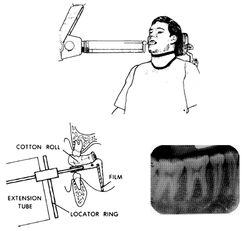
Position the instrument assembly in the patients mouth with the bicuspids centered on the film assuring that the film is parallel both vertically and horizontally. Centering the bicuspids may not be possible in patients with small mouths. Therefore, position the film in the center of the mouth as far forward as possible, touching the curvature of the lower arch (see figure 4-26). Parallel film placement is the key; it prevents dimensional distortion and overlapping. With the plastic bite-block held in place by the occlusal surfaces of the mandibular bicuspids, insert a cotton roll between the block and the maxillary teeth. Have the patient close his teeth holding the film in place. Slide the locator ring along the indicator rod bringing it close to the skin surface and align the x-ray unit extension tube with both the rod and the ring on the horizontal and vertical planes.