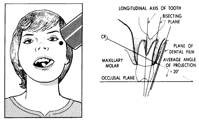
MAXILLARY MOLARS
Adjust the head as described for radiographs of maxillary teeth (refer to paragraph 4-7a). Place the film packet in the mouth so that its long axis is horizontal, the anterior border of the film is lingual to the mesial border of the second bicuspid, and the lower border of the film is parallel to and slightly below (approximately 1/4 inch) the occlusal surfaces of the molars. The upper corner of the packet may be contoured slightly but the film packet should not be bent. Adjust the tube to an average angulation of +20º. Direct the central ray straight through the interproximal spaces in the area of the second molar and perpendicular to the bisecting plane (see figure 4-10). Follow the manufacturer's instructions for all exposure times.