
POSITIONING THE PATIENT
Standard radiographic procedures include precise positioning of the patient's head as one step in placing film. The tissues to be radiographed and the x-ray beam must be in proper relationship to produce an accurate radiographic image. This is particularly important when using the bisecting angle technique. In adjusting the backrest and headrest, it is important to make the patient as comfortable as possible to minimize movement during exposure. As in photography, movement during exposure will result in a blurred image. Blurring may be greatly reduced through the use of ultra-speed film.
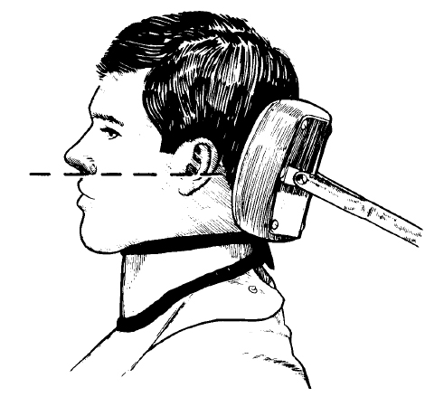
a. Head Positioning in Radiography of the Maxilla Using the Bisecting Technique. In radiography of the maxilla, the head should be positioned so that the occlusal surfaces of the maxillary teeth are in a horizontal plane (see figure 4-2). This is done by adjusting the headrest so that the median plane (sagittal plane) is vertical and a line from the ala of the nose to the tragus of the ear is horizontal.
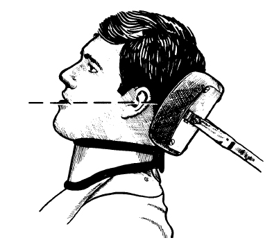
b. Head Positioning in Radiography of the Mandible Using the Bisecting Technique. In periapical radiography of the mandible, the head should be positioned so that the occlusal surfaces of the mandibular teeth will be horizontal when the mouth is opened to the position in which the radiographs are to be made (see figure 4-3). This is done by adjusting the headrest so the median plane is vertical and a line from the corner of the mouth to the tragus of the ear is horizontal.
c. Angulation. When the cone is adjusted to project the central beam upward, it will be set at a negative (-) degree angulation. When it is adjusted to project the central beam downward, it will be set at a positive (+) angulation.