
RADIATION HAZARDS AND PROTECTION
RADIATION PROTECTION
a. General. Filtration and collimation of the x-ray beam are very important safety measures. The filter and collimator (diaphragm) block the majority of the unwanted x-ray photons. As you progress through the next few paragraphs of this text, you will understand their importance. The following diagram will identify the location of these two devices (see figure 1-8).
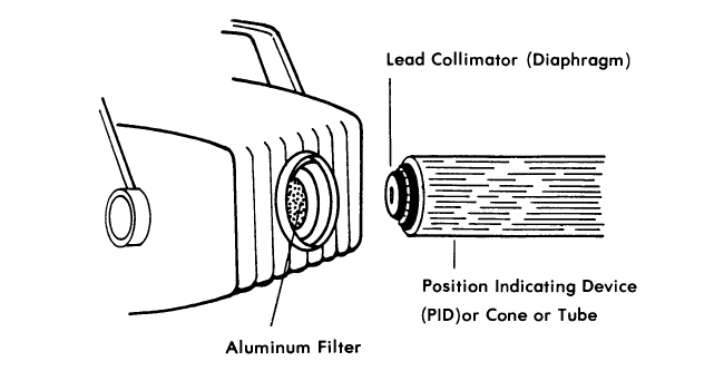
b. Filter. The aluminum filter or disk is placed in the path of the x-ray beam. It is located at the base of the cone or position indicating device (PID) just inside the metal housing. Figure 1-8 shows the location of the PID. The filter completely covers the opening where the x-ray beam emerges from the x-ray tube. The reason for the aluminum filter is to absorb the low energy, long wavelength x-rays (photons) and allow the high energy, short wavelength x-rays (photons) to pass through the filter. Filters on dental x-ray machines with over 70 kVp have a minimum thickness of 2.5 mm of aluminum. Those machines below 70 kVp have a safety standard minimum of 1.5 mm aluminum.
NOTE: The terms cone, PID, or tube are used interchangeably throughout this text. See figure 1-8.
c. Collimator. The lead diaphragm is collocated with the aluminum filter. It restricts the x-ray beam to the desired size. The diaphragm or collimator is constructed of 1/16-inch lead. Without this collimator, x-ray photons would cover a wide area of the patient's head. With the lead diaphragm or collimator in place, only the area necessary for exposure receives the primary beam. This is depicted in figure 1-7. The diagram in figure 1-8 represents an x-ray tube, cone, or PID removed to show the location of the lead diaphragm or collimator and the aluminum filter.