
DENTAL RADIOGRAPHIC FILM
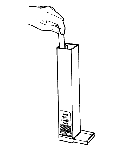
Dental radiographic film is supplied in various sizes and degrees of sensitivity, each designed for a specific purpose. Film should be stored in a cool, dry place free from chemical contamination. High temperatures, moisture, and certain chemicals will cause deterioration of the film's emulsion. Dispensers made of radiopaque metal are commonly used to provide a limited stock of periapical films. Lead-lined boxes are available for storage and protection of film used in the dental clinic (see figure 3-1). An expiration date is stamped on each film package by the manufacturer. Film should not be overstocked, but maintained in quantities that will be used before the expiration date.
a. Film Construction.
(1) An x-ray film has two parts, the cellulose acetate base and the emulsion covering it. The base is made of cellulose and is a transparent plastic that is clear or with a slightly bluish tint. The emulsion is a thin coating of a special gelatin with minute particles of a silver compound. When x-rays penetrate through soft tissue, such as the cheek or the gingiva, they also penetrate the emulsion easily, which causes the silver compound to stick to the cellulose base during processing. Therefore, the film is darker (this is known as radioluscence). When x-rays are directed to hard tissue (which is dense), such as bone, tooth enamel, and metal restorations, fewer x-rays reach the film. So, more of the silver compound is removed by processing. This results in lighter shades on the film (which is known as radiopaque).
(2) All dental x-ray films (intraoral films) have an embossed dot (a raised spot) to identify right and left. This raised dot is on the side of the film facing the x-ray tube. The purpose of the embossed dot is to assist in mounting radiographs in correct anatomical order.
b. Types of Film.
(1) Intraoral film. Periapical, bite-wing, and occlusal are three types of intraoral film used to reveal different dental structures.
(a) Periapical film is used primarily for radiographic examination of teeth and adjacent tissues to include the periapical region. The standard periapical film (Type 2) used in the Army is 1 1/4 by 1 5/8 inches, which is large enough to include a view of about three teeth. A small size periapical film (Type O) is also a standard item of issue for use in radiography of children's teeth and measures 7/8 by 1 3/8 inches.
(b) Bite-wing film is used to obtain a radiograph of the coronal two-thirds of opposing maxillary and mandibular teeth and their adjacent tissues on a single film. The film packets are provided with tabs that extend from the center of the film. When a radiograph is being made, the patient is instructed to bite down on the tab. The tab holds the film firmly in position with the lower half lying lingual to the mandibular teeth and the upper half held lingual to the maxillary teeth. Type 3 is the standard type of bite-wing film used in the Army. It measures 1 1/16 by 2 1/8 inches. When the type 3 bite-wing film is unavailable or if the dental officer requests it, the type 2 periapical film may be used to take bite-wing x-rays. However, these films would require the use of paper adapters. Type O periapical film may be used as a bite-wing film for children. These, also, would require the use of paper adapters.
(c) Occlusal film is a highly sensitive double-emulsion film supplied in packets similar to periapical film but in a size convenient for obtaining a view of the entire upper or lower arch or portions thereof. It measures 2 1/4 by 3 inches. Some packets contain two films. The first film is developed at normal time to give a detailed image of hard structures. The second film is developed in one half the normal time to reveal soft tissue images.
(2) Extraoral film. Extraoral film is used for radiographs of the jaws, facial bones, the temporomandibular joints, and other relatively large areas. This film has no embossed dot to identify right and left.
(a) Intensifying screens are used with extraoral film to intensify the effects of the exposing rays and lessen the exposure time.
(b) A cassette is constructed of rigid metal, plastic, or cardboard. It often contains intensifying screens that magnify the x-ray beam,
thus reducing exposure time. The film must be transferred to the cassette from its paper covering in the protection of the dark room.
(3) Panoramic film. Panoramic film, a type of extraoral film, is used in panoramic radiography. This film shows the entire dentition and
surrounding bone structure.