RADIOLUCENT LANDMARKS ON MAXILLARY RADIOGRAPHS
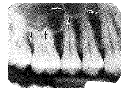
a. Maxillary Sinus. The maxillary sinus (see figure 3-21) is a very prominent radiolucent structure. It sometimes appears as overlapping lobes or a single radiolucent area with a radiopaque border. The maxillary sinus is partially seen in all periapical radiographs of the bicuspid-molar area. It occupies a large part of the body of the maxilla, varying greatly in dimension, but normally extending into the alveolar process adjacent to the apices of the posterior teeth.
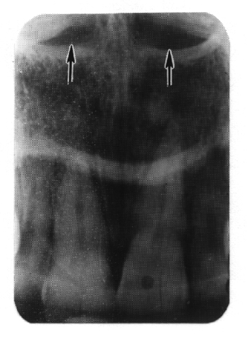
b. Incisive Foramen. The incisive foramen (see figure 3-22) is seen as a dark area located between and extending above the central incisors. In radiographs exposed from the region of the cuspid or lateral incisor, the incisive foramen may appear as a radiolucency at the apex of one of the incisors.
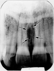
c. Median Palatal Suture. The median suture of the palate (see figure 3-23) may appear as a radiolucent line extending posteriorly from the alveolar border in the sagittal plane of the maxilla, on an anterior periapical film, or occlusal film.
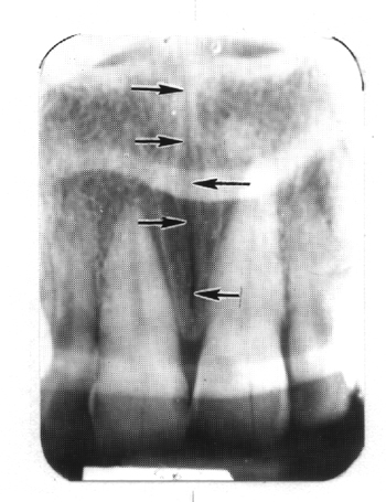
d. Nasal Fossae. In a radiograph of the maxillary central incisors, the images of the paired fossae appear as somewhat elliptical radiolucent areas of various sizes separated by a radiopaque band representing the nasal septum (see figure 3-24).