RADIOPAQUE LANDMARKS ON MAXILLARY RADIOGRAPHS
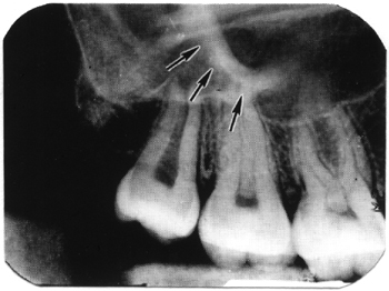
a. Maxillary Tuberosity. The maxillary tuberosity (see figure 3-25) is the convex distal inferior border of the maxilla, curving upward from the alveolar process and distal of the third molar. An extension of the maxillary sinus is occasionally seen within the maxillary tuberosity.
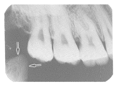
b. Coronoid Process of the Mandible. The coronoid process of the mandible (see figure 3-26) sometimes appears on maxillary molar films as a triangular opaque area located in the region of or distal to the maxillary tuberosity.
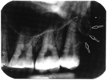
c. Zygomatic Process (Malar Bone). The zygomatic arch (see figure 3-27) commonly appears as a well-defined radiopaque area that may be superimposed over the molar roots. Additional radiographs are sometimes made at adjusted angulation to provide a better view of the molar root area.
d. Nasal Septum. The nasal septum is usually seen as a white ridge extending above and between the central incisors.